The Museum’s Biotechnology Laboratory
Sanger Sequencing, DNA Barcoding, and the Three Work Areas
Estimated reading time
5
How to read a wolf’s DNA from a single hair found in the wild: the capillary sequencer, the DNA barcoding technique, and the structure of three physically separated areas in the molecular biology laboratory of the Efisio Noussan Museum
The Efisio Noussan Regional Museum of Natural Sciences is not only the exhibition venue at Saint-Pierre Castle, but also the operational headquarters in La Salle. Among other facilities, it houses a molecular biology laboratory that performs work that is in many ways invisible yet scientifically decisive: reading the DNA of animal, plant, and fungal species present in the Aosta Valley territory, precisely identifying them, and contributing to the knowledge of Alpine biodiversity.
Established in 2013 with contributions from the Autonomous Region of Aosta Valley and the ERDF-funded project “VDNA Barcoding Research Unit,” the Museum’s biotechnology laboratory specializes in Sanger DNA sequencing and Fragment Analysis. These two techniques rely on a capillary sequencer, allowing researchers to read the sequence of nitrogenous bases and accurately estimate the size of a specific DNA fragment, respectively.
Sanger Sequencing: How It Works
The Sanger method is named after British biochemist Frederick Sanger, who developed it in the 1970s and received his second Nobel Prize in Chemistry for it in 1980. In its modern, automated version—as used in the Museum’s laboratory—the process unfolds in several distinct stages.
DNA extracted from a biological sample is first amplified using PCR (Polymerase Chain Reaction), a technique that multiplies copies millions of times, even from infinitesimal amounts of material. The amplified fragments then undergo a sequencing reaction, where molecules called dideoxynucleotides—each labeled with a different fluorescent dye corresponding to its nitrogenous base—terminate DNA synthesis, producing fragments of varying lengths.
These fragments are then separated under an electric field inside extremely thin polymer-filled capillaries via capillary electrophoresis: shorter fragments migrate faster than longer ones. As they pass a laser detector, the fluorescent dyes emit signals that software translates into an electropherogram—a sequence of colored peaks corresponding to the base sequence of the analyzed DNA.
DNA Barcoding: Identifying a Species from Its DNA
DNA barcoding uses sequencing techniques to identify biological species from trace samples. The concept, proposed in 2003 by Canadian biologist Paul Hebert, rests on a simple principle: just as a barcode identifies a product on a supermarket shelf, a short, standardized DNA sequence—corresponding to a portion of the genome that varies between species but remains identical among individuals of the same species—allows an organism to be identified even when visual observation is impossible.
For animals, the most commonly used “barcode” is a segment of the mitochondrial COI (Cytochrome c Oxidase I) gene. For plants, the chloroplast regions rbcL and matK are generally used. For fungi, the ITS region of ribosomal RNA is targeted.
Applied to the wildlife of the Aosta Valley, DNA barcoding enables non-invasive species monitoring—without capturing animals, disturbing populations, or even sighting them. A single hair found on a fence, a feather gathered on a trail, scat, or a tissue fragment is enough to identify the species.
Laboratory Structure: Three Physically Separated Areas
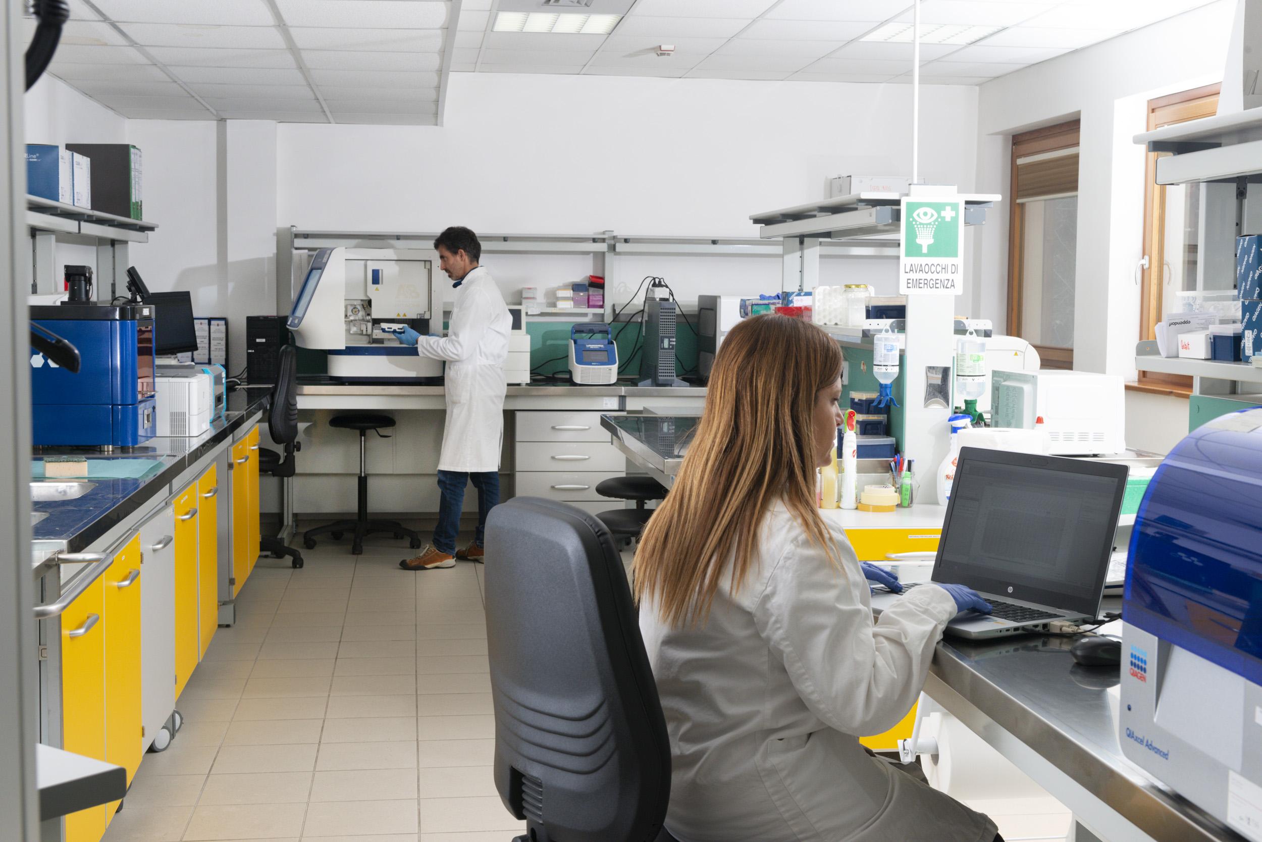
The quality of results produced by the biotechnology laboratory depends heavily on its spatial organization. The lab is structured into three physically separated areas, categorized according to the risk of DNA contamination.
NO DNA AREA: The most strictly controlled zone, where solutions and reaction mixes for PCR and Cycle Sequencing are prepared. It is equipped with a PCR hood featuring UV lamps to decontaminate work surfaces, technical and analytical balances, an autoclave, and a temperature-controlled incubator. No DNA extracts are allowed in this zone, as even minimal traces could cause contamination during subsequent amplification steps.
LOW DNA AREA: The zone for sample intake, preparation, and DNA extraction. Here, biological samples are logged, photographed, and, if necessary, observed under microscopes—the lab features both a transmission optical microscope and a stereomicroscope, both connected to a digital camera. DNA is extracted using procedures involving centrifuges, shakers, and thermomixers. A sterile laminar flow biosafety cabinet is also present for extracting DNA from samples requiring exceptionally controlled conditions, such as environmental DNA (eDNA).
HIGH DNA AREA: The zone where amplified DNA is analyzed. This area houses the PCR thermal cycler, the DNA electrophoresis station, and the capillary sequencer for Sanger sequencing and Fragment Analysis. A chemical fume hood is also available for handling toxic or hazardous reagents.
The physical separation between these three areas is not merely an optional precaution. PCR is so sensitive that it can amplify infinitesimal traces of DNA, including that of laboratory staff, if proper measures are not taken. Keeping preparation stages strictly separate from amplification stages is essential for producing scientifically reliable results.
Applications: From Wildlife to Agro-Food Traceability
The activities of the Museum’s biotechnology laboratory span from naturalistic research to enhancing local products. On the naturalistic front, the lab carries out non-invasive genetic monitoring projects for local animal species, including wolves, and has contributed to studying the biodiversity of Alpine fauna and wild flora using DNA fingerprinting techniques. On the agro-food front, it has developed protocols for the genetic characterization of local raw materials and product traceability throughout the supply chain.
Additionally, the laboratory welcomes university students for internships and experimental thesis projects (subject to agreements between universities and the host institution) and offers educational programs for secondary schools on biotechnology topics.
Photo: Pixabay